Thursday, September 5, 2013

Breast - Fibroadenoma

Fibroadenoma actually quite common in female patients.

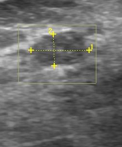
These are the ultrasound images of breast fibroadenomas


Echogenic foci is seen in fibroadenoma. Suggestive of microcalcification


Ultrasound appearance of fibroadenomas are:-
  1) Oval shape
  2) Circumscribed
  3) Homogeneous
  4) Hypoechoic
  5) Macrolobulated
  6) Smooth, thin, echogenic capsule
  7) Variable Acoustic enhancement


So What is fibroadenoma. Let's see...
- Fibroadenoma is a solid , benign breast lump.
- It contains of fibrous and glandular tissue.
- Common in adolescent and young adult population.
- Having fibroadenoma does not increase risk of developing breast            cancer.

Signs & Symptoms:-
  1) Solitary
  2) Mobile
  3) Firm
  4) Painless
  5) Size increase during pregnancy or having estrogen therapy
  6) Size atrophy after menopause, or when estrogen stimulation is
      decrease

Causes :-  Unknown
However, development of fibroadenoma probably is related to reproductive hormones. Because fibroadenoma most common in reproductive years

Others Imaging diagnosis:-
--> Mammogram : Fibroadenoma appears as Circumscribed;  Round/oval shape;  Occasionally have coarse                               calcification

Impression:
1) Mostly fibroadenoma are left in situ and follow up by ultrasound to see if it have any changes or grow.
2) Some are surgical excision.   The reasons of removal fibroadenomas:-
                                                      - Abnormal Biopsy result
                                                      - Pain or other symptom occur
                                                      - Concern about cancer
3) Biopsy recommed to confirm diagnosis.   Indication for biopsy :-
                                                                      - Size enlarged
                                                                      - Atypical findings on ultrasound
                                                                      - Lesions > 2.5cm, where no previous ultraound for                                                                                       comparison

Notes:
1) Giant fibroadenoma  --> more than 5cm size
2) Complex fibroadenoma --> Cystic changes; calcifications
3) Juventile fibroadenoma --> found in an adolescent girl


Sources:
1) Wikipaedia
2) Medline Plus
3) Mayo Clinics
4) Emedicine.medscape
5) Health guide online


Saturday, November 27, 2010

Hepatomegaly

Hepatomegaly


Introduction

  • Is liver englargement
  • The lower edge of the liver normally comes just to the lower edge of the ribs (costal margin) on the right side. 


Clinical Presentation
  • Pain 
  • Jaundice
  • Abdominal mass
  • Tenderness


Causes 
    1. Liver displacement (Palpable liver, no Hepatomegaly)
      1. Obstructive lung disease (AsthmaCOPD)
      2. Adjacent mass (Cholecystitis, abscess)
      3. Thin patient
    2. Infectious
      1. AIDS
      2. Hepatic Abscess (e.g. Amebiasis)
      3. Relapsing Fever
      4. Echinococcal cysts
      5. Schistosomiasis
      6. Kala Azar
    3. Hepatitis
      1. Alcoholic Hepatitis
      2. Autoimmune Hepatitis
      3. Viral Hepatitis
    4. Congestive Hepatomegaly (often precedes edema)
      1. Congestive Heart Failure (systemic venous congestion)
      2. Constrictive Pericarditis
      3. Tricuspid stenosis
    5. Miscellaneous
      1. Wilson's Disease
      2. Fatty Liver
      3. Cirrhosis
      4. Hemochromatosis
      5. Primary Sclerosing Cholangitis
      6. Cholestasis
      7. Mixed cryoglobulinemia
      8. Congenital hepatic fibrosis
      9. Polycystic liver disease
      10. Hurler's Syndrome
      11. Gaucher's Disease
      12. Sarcoidosis
      13. Amyloidosis
    6. Malignancy
      1. Intraabdominal malignancy
      2. Pancreatic Cancer
      3. Hepatocellular Carcinoma
    7. Toxic/Drug-related
      1. Alcoholic liver disease: acute alcoholic hepatitis and alcoholic fatty liver
      2. Drug induced hepatitis, e.g. statins,macrolidesamiodarone, paracetamol (indicates significant damage).
Causes (important n why?)
  • Alcohol abuse. Abusing alcohol may cause an enlarged liver.
  • Nonalcoholic fatty liver disease. This includes a range of conditions — such as fat accumulation in the liver followed by inflammation of the liver and the formation of fibrous tissue in the liver — which can affect people who drink little or no alcohol. 
  • Hepatitis. Hepatitis A, hepatitis B and hepatitis C are all contagious viral infections that affect your liver.
  • Heart failure. Also called congestive heart failure, this condition means your heart can't pump enough blood to meet your body's needs.
  • Liver cancer. Cancer of the liver is often metastatic cancer, which occurs when tumors from other parts of the body spread (metastasize) to the liver.
  • Infections such as mononucleosis. Mononucleosis (mono) is an infection with the Epstein-Barr virus.
  • Excessive iron in the body (hemochromatosis). This condition causes your body to absorb too much iron from the food you eat. The excess is stored in your organs, especially your liver, heart and pancreas. Sometimes the stored iron damages these organs, leading to life-threatening conditions such as cancer, heart problems and liver disease.
  • Blood disorders such as leukemia. Leukemia is cancer of the white blood cells
Risk Factors
  1. Alcohol abuse
  2. Obese 
  3. Diabetes
  4. Abnormal cholesterol levels
  5. Heart failure. Various factors increase the risk of heart failure, including high blood pressure, coronary artery disease, diabetes, kidney failure and congenital heart defects.
  6. Smoking
  7. Hepatitis A,B,C
Question to Patient

  1. Did you notice a fullness or lump in the abdomen?
  2. What other symptoms do you have?
  3. Is there any abdominal pain?
  4. Is there any yellowing of the skin (jaundice)?
  5. Is there any vomiting?
  6. Is there any unusual-colored or pale-colored stools?
  7. Have you had any fever?
  8. What medications are you taking?
  9. How much alcohol do you drink?
Ultrasound
  • Measurement at mid-clavicular line:   >15.5cm

Sunday, September 5, 2010

Liver - Sonogram

Normal Liver

Transverse scan (TS) 


Rt lower lobe of liver show homogeneous hepatic parenchymal echo pattern as isoechoic or slightly echogenic than renal parenchymal.

Rounded echogenic areas represent fibrofatty tissue surrounding ducts and vessels too small to be imaged.


Longitudinal scan (LS)

Homogeneous liver echo pattern as isoechoic or slightly less echogenic than pancreas.

Liver - Anatomy

Normal Liver 


  • Lies in upper quadrant of abdomen
  • Suspended from right hemidiaphragm
  • Have 3 lobes: (right, left, caudate lobe)
  • Right intersegmental fissure divided  RT lobe into anterior and posterior segment 
  • Left intersegmental fissure divided LF lobe into medial and lateral segment
  • Caudate lobe locate posterior of liver


Liver occupies the Rt hypochondrium, the greater part of epigastric region, & extends in varying degrees into Lf hypochondrium as far as mammary line.

Lateral segment of Lf lobe & the length of Rt lobe determine contour and shape of liver. 

Liver can described as irregular, hemispheric, or wedge shape.


Caudate lobe
  • Situated at posterior of liver
  • Posterior border: IVC
  • Anterior border: Ligamentum venosum
  •  

Abdomen and superficial structures

 By Diane M. Kawamura pg 120

Saturday, September 4, 2010

Liver - Technique

Normal Liver


Technique

  • Patient: Fasting 6 hours (so the bowel gas limited & GB is not contracted.)
  • Position: Supine, right anterior view (bec liver is tucked beneath the lower right ribs, a transducer with a small scanning face allowing an intercostal approach, is valuable.)
  • Breathing: Suspended inspiration (so able to see dome)
  • Transducer: Sagittal, transverse, coronal, subcostal oblique


To make the liver more accessible, patients need:

  • Raise the right arm above the head (to draw the rib cage upward)
  • Supine position
  • Deep breath and hold (to expand the abdomen)
Transducer/ Scanning approach:


Scanning down the left border of liver (TS)


Liver TS scan: Center of liver


*Place the transducer in the epigastrium region to the left of midline, for imaging LF hepatic border*

Liver TS scan: Right lobe


* Hold transducer in a slightly oblique position, parallel to the costal margin *


Step for Liver intercostal scan:

  1. Scan through the liver in fan-shaped pattern
  2. Slide the transducer along the intercostal space & scan through
  3. Move transducer to next intercostal space & scan through

Liver intercostal scan